The innate immune system is vital for controlling infections. However, this system is prone to being hijacked by infecting pathogens. As the host’s cellular machinery has evolved to restrict infection, so has the pathogen co-evolved to counteract cellular immune responses. While there is substantial evidence for stimulation of innate immune signaling in cells infected with L. monocytogenes, its effects on bystander cells is still largely elusive.
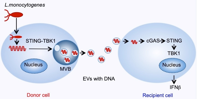
This study uncovers a role for DNA loaded extracellular vesicles released from infected cells that induce a cGAS-STING-TBK1 mediated interferon (IFN) response in bystander cells. The activation of interferon response in T-cells and resulting inhibition of T-cell proliferation sets the stage for bacterial dissemination.
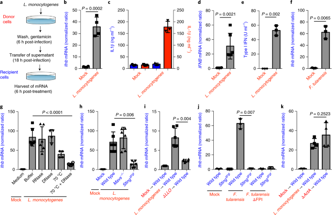
This study was prompted by a finding that cell culture media of infected cells was able to induce an interferon response in bystander cells. This indicated the presence of an entity that was not the infective pathogen itself that was capable of relaying danger signals leading to IFN induction in uninfected cells. These findings immediately directed our focus toward the usual suspects. Was the observed bystander activation an effect of cGAMP-mediated response, or perhaps a bacteria-derived product?
Surprisingly, where wild type cells stimulated with the supernatant of infected cells could induce IFN, cGAS deficient cells could not. This proved against the model of cGAMP or cyclic di-AMP acting as the IFN stimulator and prompted us to consider the stimulant to be pathogenic DNA; probably hitchhiking within vesicles to be introduced into bystander cells.
These observations as tested by our collaborators were not limited to Listeria alone, as experiments with Gram-negative Francisella tularensis and Legionella pneumophila displayed a similar phenotype.
During the period of this study, reports of DNA in vesicles from cancer cell lines were beginning to emerge. However, there was little evidence of pathogenic DNA being packaged in vesicles. This compelled us to verify the presence of bacterial DNA in vesicles through a plethora of different techniques (Confocal microscopy, Atomic Force Microscopy, NGS) and it was evident that bacterial DNA had made itself available to vesicles probably by intersecting the vesicle trafficking pathway. However, that this phenomenon seemed to depend on STING and TBK1 came as a surprise to us.
Since STING was dispensable to the production of CD81+ CD63+ vesicles, and vesicles from STING deficient cells could not induce an interferon response, we dissected further into the role of STING in sorting DNA into vesicles. Preliminary investigations to compare vesicular components between Wild type and STING deficient cells using AFM revealed the absence of DNA in EVs from STING deficient cells and genome mapping revealed that the entire bacterial genome was represented within vesicles with no detectable preference for specific areas of its genome.
Owing to the role for TBK1 dependent phosphorylation, we reached out to our collaborator in Munich to ascertain phospho targets of TBK1 to identify proteins of interest that could be involved in the sorting pathway. Mass spec data revealed MVB12b, a protein involved in the ESCRT pathway. A few CRISPR knock-out assays and several functional studies later, we were faced with the thrilling certainty that phosphorylated MVB12b was essential for DNA sorting into vesicles.
We are only beginning to uncover the mechanism of how non-self DNA might be recognized and encapsulated within vesicles. While one can appreciate the ways pathogens subvert cellular pathways to a pro-pathogen stance, the exact mechanisms for the same remain largely unknown.
We hope to understand what leads to the recognition of DNA, how it is cleaved and how it is packaged. While in future we hope to be able to answer more of these questions; for now, we are grateful for the assistance of our collaborators as well as our reviewers, who have (mostly) been fair in their comments.


Please sign in or register for FREE
If you are a registered user on Research Communities by Springer Nature, please sign in