
The global impact of the COVID-19 pandemic has changed modern day life as we know it, with long standing healthcare, social, and economic implications. Global vaccination programmes are currently helping to slow down the spread of this disease, yet emerging variants and the dissemination to vulnerable populations pose a continuous public health challenge. As the virus spread across the globe in early 2020, national lockdowns were imposed and everyday life and research were put on hold. A few months prior, the Izar laboratory had moved from Boston to New York City. We had set up shop to pursue our studies of therapy resistance in cancer with a particular interest in cancer cell intrinsic mechanisms shaping the (tissue) response to immunotherapy (Jerby-Arnon et al., 2018; Frangieh et al., 2021). Suddenly, we found ourselves at the epicentre of a raging pandemic. Ambulance sirens blaring around the clock in the otherwise empty streets of Manhattan and the other boroughs, and some lab members were stuck outside the US as the once hyperconnected world had become a dangerous place to travel.
While laboratories worldwide focused their efforts on understanding how the virus operates and how the body responds to SARS-CoV-2, the local medical research community at Columbia University and other institutions implemented a bio-banking program to promote rapid translation of basic science to benefit patients. We leveraged our experience in studying complex cancer tissues using single-cell genomics and set out to study the cellular response to SARS-CoV-2 on a tissue level. To this end, we implemented a single-nuclei RNA sequencing protocol on snap frozen specimens obtained during rapid-autopsy of COVID-19 decedents.
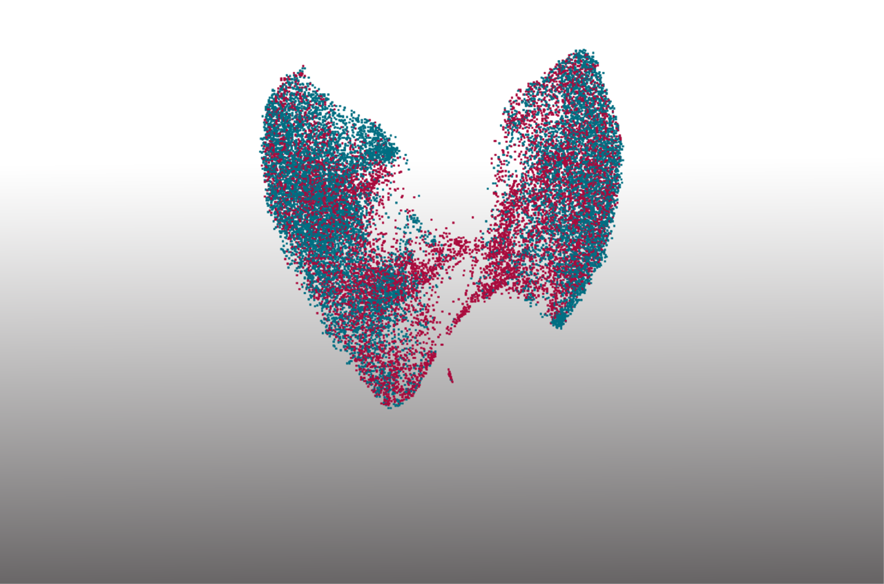
This work was the epitome of team science. During a rapid conception phase, we assembled a team of wet- and dry-lab scientists, each one bringing their unique expertise from different fields, and got to work. After optimizing each step of the protocol to work on non-infectious control tissue (which requires some modifications compared to cancer tissue) we set up a plan with the goal to analyse a range of patient samples covering different disease durations. From experience with surgical specimens, we knew that RNA-quality is inversely correlated to the time a tissue has been taken out of the body, so the most important inclusion criterium for our samples was a short post-mortem interval. Fast-forward to less than a month later, we started analysing the first data from COVID-19 lung tissue and compared the cell type composition and cell states to a set of non-infected pre-pandemic control samples, which we had processed using the same methodology. Our team of analysts and experimentalists achieved an overview of the cellular landscape and started exploring into COVID-19-specific changes using our profiling data and orthogonal experimental approaches for validation. The over-arching defining feature of COVID-19 lungs was their highly inflamed state, leading to extensive pulmonary damage, arising via the concerted actions of multiple aberrant cell responses. For instance, we observed heightened infiltration by aberrantly activated monocyte-derived macrophages and alveolar macrophages, characterised by impaired efferocytosis (clearance of apoptotic cells) that contributes to sustained inflammation during lung regeneration.
When analysing the expression features of the alveolar epithelial cells that make up the alveoli and mediate gas exchange, we noted that alveolar type 2 (AT2) cells and alveolar type 1 (AT1) cells in COVID-19 donor samples showed a transcriptional cell state which was distinct from their control counterparts and projected in between the two healthy populations. We hypothesised that the AT2 progenitor cells were differentiating in an attempt to initiate lung regeneration but failed to achieve a full transition to AT1 cells. Serendipitously, three independent mechanistic studies on lung regeneration in animal models and chronic inflammation were published around the same time as our initial observation and converged upon a cell state termed “damage-associated transient progenitors” (DATP), “alveolar differentiation intermediate” (ADI), or “pre-AT1 transitional cell state” (PATS), which was linked to increased IL1β signaling and described precisely what we observed in COVID-19 lungs. Scoring signatures derived from these publications revealed a strong signal for these cell states in our data, suggesting that, despite attempting to initiate a lung regeneration program, these stalled AT2 transitioning cells fail to undergo differentiation and become locked in an intermediary cell state which hampers regeneration in COVID-19-infected lungs.
While lung regeneration is clearly impaired in COVID-19 lungs, we observed ectopic expression of tuft cells in the parenchyma of COVID-19 lungs that was absent in control lungs, suggesting that these cells are being recruited to the lungs in response to this extensive alveolar epithelial damage in an apparent last-ditch attempt to elicit a lung regeneration programme. Further compounding this sustained pulmonary damage, we also observed a significant expansion of CTHRC1+ pathological fibroblasts in COVID-19 lungs contributing to rapid pulmonary fibrosis. COVID-19 lungs were characterised by heightened pro-inflammatory monocyte/macrophage-derived IL1β and epithelial cell-derived IL6. While IL6 was increased in COVID-19, it was not unique to this disease when compared to other pulmonary infections, and thus represents a non-specific marker of inflammation from infection. IL1β, however, was significantly enriched in COVID-19, suggesting that this cytokine uniquely contributes to the pathobiology of COVID-19 and may aid in sustaining the DATP state. Our initial target analysis has nominated several druggable pathways and targets in COVID-19-specific cell states, and we hope that this resource is of value to the community in the pursuit of disease modifying treatments for COVID-19 and similar diseases.
Early in our work we connected to our colleagues in Boston who pursued a similar effort across the hospitals in the Boston metro area. While the scope of the projects differed (the Boston group had set out to generate a multi-organ analysis), these studies showed promise to be complementary in nature. While the teams worked independently to dissect the fatal biology of lethal COVID-19 captured in each cohort, we ensured harmonisation of metadata and reporting to facilitate future analysis of all data. Our work, therefore, goes hand-in-hand with the companion manuscript by Delorey et al. and helps establish the single-cell landscape evoked by SARS-CoV-2 infection. The paper has been published in Nature online on April 29, 2021 and appears in print in the July 1st edition.


Please sign in or register for FREE
If you are a registered user on Research Communities by Springer Nature, please sign in